

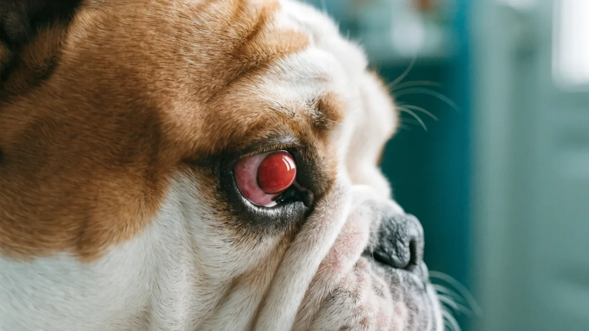
When Mr. Henderson rushed his six-month-old French Bulldog, Winslow, into my clinic on a Tuesday morning, he was practically shaking. “He was fine last night,” he said, holding the squirming pup up to my exam light, “and now he’s got this horror-movie thing hanging out of his eye.” What Mr. Henderson was looking at was a bright red, swollen mass bulging from the inner corner of Winslow’s left eye — about the size of a small cherry. He was convinced it was a tumor. I see this reaction all the time. But what Winslow had is one of the most recognizable, and fixable, eye conditions in veterinary medicine: a prolapsed gland of the third eyelid, affectionately known as “cherry eye.” And almost without fail, it’s dogs who walk through my door with it, not cats.
What Exactly Is Cherry Eye?
To understand cherry eye, you first need to know about a structure most people don’t realize their pet even has: the third eyelid, or nictitating membrane. Both dogs and cats have one. It’s a thin fold of tissue tucked away in the inner corner of each eye, near the nose. You usually only see it when your pet is sleepy, slightly sedated, or if you gently press on the eyeball through the closed lid (though I don’t recommend making a habit of that).
The third eyelid isn’t just a passive flap. It serves real purposes: it acts as a windshield wiper for debris, produces a portion of the tear film that keeps the eye lubricated, and houses a lymphoid structure that helps fight off local infections. Crucially, it also contains a tear-producing gland — and this is where the trouble starts.
In cherry eye, the ligament and connective tissue that are supposed to hold that tear gland snugly in place behind the third eyelid simply give way. The gland flips up and out of its normal pocket, protruding into view. Once exposed to air and irritation, it swells, turns bright red, and looks alarming. That red mass is not a tumor. It’s not an infection. It’s a tear gland in the wrong zip code.
Why Dogs Get It and Cats (Almost Never) Do
This is the question I get asked constantly. If both species have a third eyelid with the same gland, why is my waiting room full of dogs with cherry eye and not cats?
The Genetics of Weak Connective Tissue
The answer lies largely in genetics and selective breeding. Cherry eye is considered an inherited condition, and it overwhelmingly affects specific dog breeds — particularly those with short muzzles, prominent eyes, or loose facial skin. Bulldogs, Cocker Spaniels, Beagles, Lhasa Apsos, Shih Tzus, and Bloodhounds are all on the frequent-flyer list. In these breeds, the connective tissue anchoring the tear gland is simply weaker, likely because generations of selective breeding for head shape and facial structure have altered the anatomy in ways that compromise that little pocket of tissue.
I’ve probably seen more cherry eye cases in English Bulldogs alone than in every cat breed combined over my entire career. Their wide-set, prominent eyes and shallow eye sockets create the perfect storm.
Feline Anatomy: Built to Last
Cats, on the other hand, rarely develop cherry eye — and when they do, it’s usually noteworthy enough to warrant a case discussion among colleagues. The connective tissue anchoring their third eyelid gland appears to be anatomically stronger and more securely fastened. Their eye structure is also different: cats tend to have deeper-set eyes with less of that exaggerated facial conformation that predisposes certain dogs to problems.
That said, I want to be clear that “rare” is not the same as “impossible.” Cats can get cherry eye. It’s been documented in Burmese and Persian cats in particular, which makes sense when you consider those breeds have some of the same brachycephalic (short-skulled) features as bulldogs and pugs. But for the average domestic shorthair cat, the risk is vanishingly small.
Why That Red Mass Matters More Than You’d Think
Pet owners sometimes ask me if cherry eye is just a cosmetic issue. It’s an understandable question — after all, the dog doesn’t seem to be in excruciating pain, and often they’re still eating, playing, and acting completely normal. But leaving a prolapsed gland untreated is a mistake for several reasons.
First, the exposed gland becomes chronically irritated. Every blink drags the eyelid across swollen tissue. Dust, debris, and environmental irritants settle on a surface that was never designed to be exposed. Over time, this inflammation can lead to corneal ulcers — painful scratches or erosions on the surface of the eye itself that can become infected and threaten vision.
Second — and this is the part I really emphasize to owners — that gland produces a significant portion of your dog’s tear film. In some breeds, it accounts for up to 30-40% of aqueous tear production. If you lose that gland, you risk developing keratoconjunctivitis sicca, commonly called “dry eye,” which is a lifelong condition requiring daily medication and potentially leading to corneal scarring, pigmentation, and even blindness. I always tell my clients: that little red blob is doing important work. We want to save it, not toss it.
The Surgical Debate: Tuck, Don’t Remove
Historically, the treatment for cherry eye was simple: cut the gland out. Problem solved, swelling gone, dog looks normal again. And for decades, that’s exactly what many veterinarians did. But as follow-up data accumulated, a troubling pattern emerged. Dogs who’d had their gland removed were developing dry eye at significantly higher rates later in life. We now understand why — we’d removed a major tear producer and expected the eye to compensate. Often, it couldn’t.
Today, the gold standard in veterinary medicine is gland repositioning, not removal. The procedure involves creating a small pocket or fold in the tissue of the third eyelid and using fine dissolving stitches to tuck the gland back into its proper position. There are several techniques — the “pocket technique” and the “anchor technique” are two of the most common — but the principle is the same: preserve the gland, restore normal anatomy, and maintain tear production.
I’ll be honest with you: I’ve changed my mind on this over the years. Early in my career, when surgical techniques were less refined, I sometimes advised owners of older dogs that removal was acceptable if repositioning failed. The recurrence rates with older methods could be frustrating. But modern surgical approaches have gotten much better, and the long-term data on dry eye is compelling enough that I now recommend preservation every single time, even if it means a second surgery.
That said, recurrence does happen. If the tissue isn’t tightened adequately during surgery, or if the connective tissue is particularly weak — as it often is in Bulldogs — the gland can pop back out. I always warn owners that there’s a 10-20% recurrence rate depending on the breed and technique used. A second surgery is usually successful.
What to Expect After Surgery
Post-operative care is usually straightforward. Your dog will go home with an Elizabethan collar (the “cone of shame”) to prevent pawing at the eye, along with antibiotic and anti-inflammatory eye drops or ointments. Some swelling and redness are completely normal as the dissolving stitches do their work and the tissue heals. I always tell owners: it might look worse for a few days before it looks better. That’s the body doing its job.
What’s not normal is green or yellow discharge, significant squinting, pawing at the eye despite the cone, or a return of that telltale red bulge. Any of those warrant a follow-up call.
I saw Winslow, Mr. Henderson’s French Bulldog, back two weeks after his pocket surgery. The swelling had resolved. The gland was sitting where it belonged, tucked neatly behind the third eyelid. Mr. Henderson was relieved. “So he’s not going to be a horror movie anymore?” he asked. I laughed. “No,” I said. “Just a normal, slightly gassy French Bulldog.”
The Takeaway for Pet Owners
Cherry eye looks scarier than it is, but it’s not something to ignore or treat with home remedies. I’ve had well-meaning owners try gentle warm compresses, herbal eye washes, or even attempts to push the gland back manually. While I understand the impulse to try something at home — especially when your pet looks distressed — none of these approaches address the underlying anatomical problem, and some can cause more irritation or even introduce infection. If you see that red swelling, call your vet. Early surgical intervention gives the best outcomes and the lowest recurrence rates.
And beyond treatment, there’s a bigger picture worth considering. Conditions like cherry eye are a direct consequence of human-driven selective breeding. We’ve shaped dogs into forms we find appealing — flat faces, wide-set eyes, skin folds — and in doing so, we’ve sometimes compromised their health. The breeds most prone to cherry eye are also prone to breathing difficulties, dental crowding, and a host of other structural issues. Choosing a reputable breeder who prioritizes health over extreme conformation, or considering adoption of mixed-breed dogs with more moderate anatomy, is one of the most meaningful ways to reduce the prevalence of these inherited conditions. Every pet deserves eyes that work the way nature intended — and the more we understand about conditions like cherry eye, the better equipped we are to advocate for the animals who depend on us.