

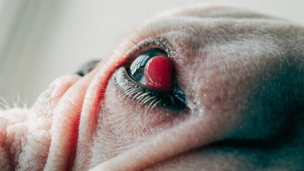
Not long ago, a frantic client rushed a six-month-old French Bulldog named Buster into my clinic. She was convinced Buster had somehow scratched his eye open overnight because a bright red, swollen mass had suddenly appeared in the corner of his lid. Meanwhile, just last week, I had a phone call from a concerned owner whose eight-month-old rescue kitten, Cleo, looked like she was wearing fleshy, translucent contact lenses. Both of these pet parents were describing issues with the exact same piece of anatomy: the nictitating membrane, or the “third eyelid.”
It’s an anatomical feature we humans lost through evolution, but dogs and cats rely on it every single day. And when something goes wrong with it, the results can look incredibly dramatic. Whether you’re dealing with a classic dog cherry eye or the peculiar feline phenomenon known as Haw’s syndrome, understanding what that third eyelid actually does is the first step toward helping your pet.
The Hidden Shield: What is the Nictitating Membrane?
If you’ve ever seen your dog or cat wake up from a deep sleep and noticed a whitish or pinkish film sliding across their eye before they blink it away, you’ve witnessed the nictitating membrane in action. Tucked snugly in the inner corner of the eye, this cat third eyelid (which dogs have, too) acts as a built-in windshield wiper.
Its job is incredibly important. It acts as a physical shield, sweeping across the eye to clear away dust, pollen, and debris. It also helps distribute the tear film evenly across the surface of the eye. But it doesn’t just spread tears; for dogs, it actually manufactures them. The gland of the third eyelid is responsible for producing a significant portion of the eye’s watery, protective tear film. When everything is working correctly, this membrane stays neatly tucked away, doing its job in the background. But when the anatomy fails or the nervous system gets confused, it becomes impossible to ignore.
When Things Go Wrong in Dogs: The Classic Cherry Eye
While cats occasionally get them, a prolapsed gland—universally known as dog cherry eye—is overwhelmingly a canine problem. It happens when the tiny connective tissues that hold the tear-producing gland in place simply give way. Once that anchor breaks, the gland pops out of its hiding spot and flips upward, sitting exposed in the corner of the eye like a small, angry cherry.
This mechanical failure is especially common in young dogs under the age of two. If you have a flat-faced, brachycephalic breed—think French Bulldogs like Buster, Pugs, Cane Corsos, or Lhasa Apsos—they are genetically predisposed to this issue. Why? Because their shallow eye sockets simply don’t hold the eye, and the structures behind it, quite as securely as they should.
The Great Surgical Debate
When that red mass appears, pet owners usually notice their dog pawing at their face, squinting, or developing a thick discharge. Naturally, they want it gone immediately.
Years ago, when I first started in practice, it wasn’t entirely uncommon for veterinarians to simply surgically remove that prolapsed gland. It got the red bump out of the way, and the dog went home looking normal. But,[1] we realized we were making a massive mistake. I’ve definitely changed my mind on how I approach this condition based on how the research developed.
Because that gland produces up to 50% of the eye’s tear film, removing it frequently led to a condition called Keratoconjunctivitis sicca, widely known as “dry eye.” Dry eye is a painful, chronic condition that requires daily, lifelong medication to prevent the cornea from ulcerating. Today, the.[2] Instead of cutting the gland out, we carefully tack it back down into its proper pocket using a buried suture technique. It preserves the tear production and resolves the irritation.
The Feline Twist: Haw’s Syndrome in Cats
If dogs suffer from a mechanical failure, cats suffer from a neurological one. When I got that call about Cleo the rescue kitten, her owner described exactly what happens in Haw’s syndrome. Instead of a red, swollen cherry, both of Cleo’s third eyelids had slowly protruded up across the bottom half of her eyes, accompanied by a slight drooping of her upper lids (a medical term we call ptosis).
It looks terrifying to an owner. You might assume your cat is going blind. But surprisingly, in cases of Haw’s syndrome, a cat’s vision usually remains completely normal. If you watch them navigate a room, they won’t bump into furniture; they just look like they’re constantly wearing a fleshy, foggy veil.
The Gut-Eye Connection
The most fascinating thing about Haw’s syndrome is that it isn’t strictly an eye issue at all..[3] When Cleo came into the clinic, her owners mentioned she had been dealing with intermittent diarrhea for a few days prior to her eyes changing.
We frequently see this in young cats, especially those from shelters or rescue environments. It’s often triggered by a viral infection—like feline herpesvirus or calicivirus—or sometimes by a heavy intestinal parasite load. The exact physiological pathway of how gut inflammation causes the third eyelid to elevate is still a bit of a mystery, but we know there’s a profound link between the enteric nervous system (the gut) and the cranial nerves that control the muscles of the eye.
Treatment and Management
Managing these two conditions requires completely different playbooks.
For a dog cherry eye, the solution is almost always surgical. While you wait for your procedure, your vet might prescribe a topical anti-inflammatory drop to reduce the swelling and prevent the gland from drying out. I completely understand the impulse to try home remedies when you see a scary-looking mass on your dog’s eye, but please don’t apply human eye drops or herbal compresses without consulting a professional. The cornea is incredibly delicate, and the wrong over-the-counter drop can do more harm than good. (And always consult your vet for exact dosages and treatments before applying anything).
For cats with Haw’s syndrome, the approach is far less invasive. Because the root cause is usually gastrointestinal, we treat the gut. Recently, [4] reinforced what many of us in clinical practice have seen firsthand: Haw’s syndrome can often be completely resolved simply by putting the cat on a specific, highly digestible gastrointestinal diet or treating the underlying parasite/viral issue. Once the gut heals and the diarrhea resolves, the nervous system calms down, and those protruding third eyelids naturally slide back into their hiding spots within a few weeks.
Whether you are raising a flat-faced puppy or a tiny rescue kitten, taking care of our domestic companions means paying close attention to the subtle signals their bodies send us. By understanding the delicate mechanics of the eye and the surprising connections between the gut and the nervous system, we can ensure our pets stay comfortable, pain-free, and enjoying the world around them with clear, bright eyes.
Sources
- as veterinary ophthalmology evolved — vet.cornell.edu
- universal standard of care is surgical repositioning — eye-vet.co.uk
- It is almost always accompanied by gastrointestinal (GI) distress — veterinarypartner.vin.com
- a fascinating 2025 study by B. Fruchter and colleagues — ncbi.nlm.nih.gov