

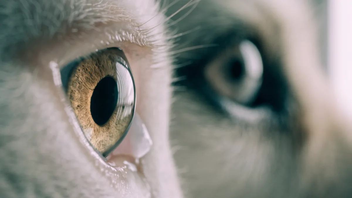
Just last week, I got a frantic text from a client. Her senior tabby cat, Whiskers, had been sleeping on the windowsill in a patch of afternoon sun. When he woke up and opened his eyes, she saw a strange, whitish film flash across his eye before it quickly disappeared. She was convinced he was going blind or had suffered a stroke. I had her bring him right in, but I already had a pretty good idea of what was going on. Whiskers was perfectly healthy. He had just given us a fleeting glimpse of his nictitating membrane, commonly known as the third eyelid.
If you’ve ever caught a sudden, spooky flash of white or pink in the corner of your dog or cat’s eye, you’ve witnessed this hidden shield in action. Most of the time, we don’t even know it’s there. But understanding what this membrane does—and recognizing when it’s trying to tell you something—is one of the most fascinating parts of caring for our pets.
More Than Just a Blink: What Is the Third Eyelid?
The scientific term for this structure is the nictitating membrane, which comes from the Latin word nictare, meaning “to blink.” If you look closely at the inner corner of your pet’s eye near the nose (the medial canthus), you might see a tiny fold of tissue. That’s the leading edge of a whole extra eyelid tucked neatly underneath the lower lid.
Humans don’t have a functional third eyelid. We only have a tiny, useless vestigial remnant in the inner corner of our eyes called the plica semilunaris. But for our pets, it’s a highly specialized piece of biological engineering.
Unlike the upper and lower eyelids, which move up and down, the third eyelid sweeps horizontally across the eye. Think of it like a microscopic windshield wiper. When a dog or cat bounds through tall grass, digs in the dirt, or stalks through brush, this membrane sweeps across the cornea to clear away dust, pollen, and debris. It even helps distribute the tear film evenly over the eye, keeping it lubricated and protected from scrapes.
The Eye’s Built-In Immune System
While it’s great at swiping away physical debris, the third eyelid is also deeply involved in your pet’s ocular health. It isn’t just a piece of tough tissue; it houses a highly active gland that produces a massive portion of the eye’s tear film. If you’re looking for a specific feature when evaluating your pet’s eye health, you want to know that this gland is functioning properly to keep the eye bright and moist.
Beyond simple lubrication, the third eyelid contains lymphoid tissue. This tissue acts like a tiny military base, producing antibodies that fight off bacterial and viral infections right at the surface of the eye. Every time that membrane sweeps across the cornea, it’s not just cleaning the lens—it’s delivering a fresh coat of protective immune cells. It’s a brilliant evolutionary adaptation that keeps animals healthy in dirty, wild environments.
When the Hidden Shield Makes an Appearance
Under normal, healthy circumstances, the third eyelid should be nearly invisible. The only time you should really see it is when your pet is deeply relaxed or falling asleep. This is especially common in cats. I often see what I call the “cat sleep” phase in my clinic—Whiskers will be sitting upright on the exam table, fast asleep, and his eyelids will slowly drift apart, leaving those pale membranes partially exposed. As soon as he hears the treat bag crinkle, they snap back out of sight.
However, if you suddenly see that membrane covering a third to a half of your pet’s eye while they are wide awake and alert, it’s almost always a medical red flag. Here are the three most common culprits I see in practice:
1. “Cherry Eye” (Gland Prolapse)
One of the most startling conditions I treat is affectionately known as “Cherry Eye.” This happens when the gland of the third eyelid prolapses, essentially popping out of its normal position and swelling into a bright red, angry mass in the corner of the eye.
I see this most often in young dogs, particularly those with short snouts and shallow eye sockets. I’ll never forget treating an eight-month-old French Bulldog named Barnaby. His owners rushed him in, absolutely convinced he had a tumor growing in his eye. Fortunately, it was just a prolapsed gland. It looks scary, but it’s rarely a true emergency.
Historically, the treatment for Cherry Eye was to simply surgically remove the swollen gland. I used to do this early in my career, but I’ve changed my mind entirely as veterinary research evolved. Removing that gland destroys up to 50% of the eye’s tear-producing capability, inevitably leading to a painful, lifelong condition called Dry Eye (Keratoconjunctivitis Sicca). The modern, universally accepted standard is the “tuck” or pocket technique. Using advanced microsurgery, we create a tiny fold of tissue to securely tuck the gland back into its proper place, preserving the eye’s natural moisture and keeping scarring to an absolute minimum.
2. Haw Syndrome (Systemic Warning Signs)
Sometimes, both third eyelids will protrude simultaneously, covering the bottom half of both eyes. This is known as Haw Syndrome. Surprisingly, it rarely means there is a primary eye problem.
More often, the third eyelids pop up because the pet is experiencing severe gastrointestinal distress, dehydration, or a systemic illness. I recently treated a three-year-old Beagle named Daisy who had been battling a nasty intestinal virus. When her dehydration hit its peak, her body naturally sacrificed some of the tone in her facial muscles, causing those membranes to slide up. Once we got her rehydrated with fluids and her stomach settled, the eyelids slid right back down to their normal hiding spots. The eyes were simply acting as a billboard for what was happening in her gut.
3. Corneal Ulcers and Pain
When a pet feels a sharp, stabbing pain in their eye, their brain tells the third eyelid to deploy as a protective shield. If a dog or cat has a scratch on their cornea (a corneal ulcer), or if they have a piece of grit or a grass awn trapped under their lid, the third eyelid will aggressively sweep across the eye to try and dislodge it, eventually getting stuck in the “up” position.
This is usually accompanied by excessive squinting, tearing, and pawing at the face. If you notice these symptoms, it’s a true veterinary emergency. Corneal ulcers can deepen rapidly, and we need to apply specific medicated drops—often containing antibiotics and pain relief—to help the eye heal before permanent scarring occurs. Always consult your veterinarian before administering any eye drops, as giving the wrong medication can worsen a scratch.
The Flat-Faced Dilemma
Right now, one of the biggest conversations in veterinary medicine revolves around brachycephalic, or flat-faced, breeds like Pugs, Shih Tzus, and Persian cats. Because of their genetics, these pets have extremely shallow eye sockets. Their eyes practically bulge from their skulls, which pulls the third eyelids into a much more exposed, tight position.
This anatomical struggle means flat-faced pets are incredibly prone to third eyelid issues. Their glands prolapse easily, their eyes dry out faster because the bulging surface area evaporates moisture, and they are highly susceptible to corneal injuries because their eyes simply don’t have the bony protection that other breeds do. If you share your home with a flat-faced companion, keeping up with their annual eye exams and watching for dry, red, or squinting eyes is critical.
Takeaway
The nictitating membrane is a marvel of evolutionary design—a built-in windshield wiper, immune booster, and protective shield all rolled into one. While it might give us a brief scare when it flashes across our pet’s eye during a Sunday afternoon nap, it’s ultimately there to keep them safe, clear-eyed, and ready to chase the next squirrel.
Take a moment today to look closely at your dog or cat’s eyes in good, natural lighting. Notice the smooth, glossy surface and the sharp, clean corners. Knowing what your pet’s eyes look like when they are perfectly healthy is the best way to catch a problem early. If you ever see a red mass, persistent squinting, or a stubborn film that refuses to retreat, don’t panic—just give your vet a call. Most of the time, with a little medical intervention, we can get that hidden shield right back to work.