

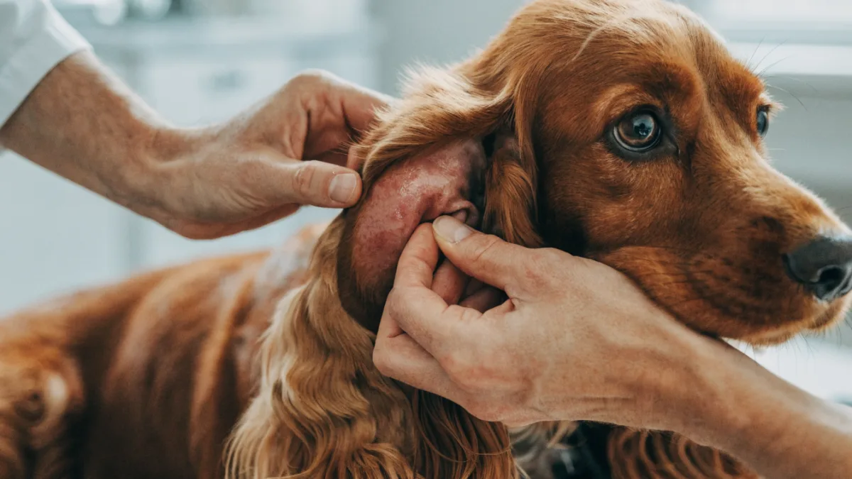
The phone calls usually sound the same. “Doctor Mitchell, Buster’s ear just blew up overnight — it looks like a balloon!” When I ask them to describe what happened, the story almost always includes a day or two of frantic head-shaking, some pawing at the ear, and then suddenly, a swollen ear flap that wasn’t there when everyone went to bed. The first time you see it, it’s genuinely alarming. That fat, fluid-filled pouch on your dog’s ear flap is called an aural hematoma, and while it looks dramatic, understanding what’s happening beneath the surface takes much of the fear out of the situation. Let me walk you through why this happens, why certain pets are predisposed, and what we can do about it.
The Cartilage Sandwich: Understanding Ear Flap Anatomy
To understand why a swollen ear flap in pets happens so easily, you need to picture what the ear flap — technically called the pinna — actually is. Think of it as a sandwich. The “bread” on either side is a layer of skin, and the “filling” in the middle is a stiff piece of cartilage that gives the ear its shape. That cartilage is covered by a blood-rich layer of skin that’s packed with delicate blood vessels.
Here’s where things go wrong. When a dog or cat shakes their head violently — the kind of whipping, full-body head shake that makes their ears slap against their skull — those fragile blood vessels between the skin and cartilage can rupture. It doesn’t take much. Once a vessel breaks, blood starts pooling in that “sandwich” space, separating the skin from the cartilage. That pooling blood is what creates the swollen, fluid-filled pocket you’re feeling. The medical term for this collection of blood is a hematoma, and when it happens on the ear, we call it an aural hematoma.
I remember a case from last spring — a six-year-old Cocker Spaniel named Rosie whose owner, Karen, brought her in on a Saturday morning. Rosie’s right ear was swollen to nearly twice its normal thickness, warm to the touch, and clearly tender. Karen was convinced something had bitten her overnight. In reality, Rosie had been dealing with a low-grade yeast infection in that ear for about a week, the itching had escalated, and the violent head-shaking had done the rest. This domino effect — underlying irritation leads to itching, itching leads to shaking, shaking leads to vessel rupture — is the story behind almost every dog aural hematoma I’ve ever treated.
Why Floppy-Eared Dogs Bear the Brunt
If you own a Golden Retriever, Cocker Spaniel, Basset Hound, or Labrador, you’re probably already familiar with the recurring ear infection cycle. There’s a structural reason for this. Floppy ears create a warm, dark, and moist environment inside the ear canal — basically a perfect incubator for yeast and bacteria. Dogs with upright, prick ears (like German Shepherds or Huskies) get more airflow and tend to have fewer ear canal infections.
That trapped moisture is why floppy-eared breeds end up at the vet with ear issues so frequently. The infection causes inflammation and itching — what we call pruritus in medical terms — and the dog responds the only way they can: by shaking their head and scratching. The heavier the ear flap, the more forceful the shaking, and the more likely those delicate vessels are to burst.
When Rosie’s mom asked me why this happened “out of nowhere,” I explained that it rarely does. The ear infection had probably been simmering for days. By the time the hematoma appeared, the infection was well-established and the head-shaking had reached a level of intensity that ruptured the vessels. Treating the hematoma without addressing the infection underneath would be like bailing water from a boat without plugging the hole.
Warning Signs to Watch For
Catching the cycle early can sometimes prevent the hematoma from forming in the first place. Watch for:
- Excessive head tilting or turning to one side
- Violent, whipping head shakes — especially if they’re repetitive
- Pawing, scratching, or rubbing the ear against furniture
- Redness, odor, or discharge visible in the ear canal
- A sudden “fat” or thickened appearance to the ear flap
- Whining, yelping, or pulling away when the ear is touched
If you’re seeing several of these together, don’t wait for the ear to swell. That’s the window where a quick ear cleaning and anti-inflammatory treatment might head off the whole cascade.
The Feline Connection: Ear Mites and Cauliflower Ear
Cats get aural hematomas too, though the trigger is often different. While dogs typically develop them secondary to bacterial or yeast ear infections, cats are more likely to end up with a swollen ear flap because of ear mites — specifically, Otodectes cynotis, tiny parasitic mites that live in the ear canal and cause intense, maddening itchiness.
Ear mite infestations are especially common in kittens, outdoor cats, and cats from multi-cat households or shelter environments. The mites themselves are barely visible to the naked eye, but they leave behind a characteristic dark, crumbly discharge that looks almost like coffee grounds. The itching they cause is severe — I’ve seen cats scratch their ears raw and shake their heads so aggressively that they ruptured vessels within hours.
A few years ago, I treated a two-year-old barn cat named Olive who came in with a swollen ear flap that her owner initially thought was an abscess from a fight. On examination, both ear canals were packed with the classic coffee-ground discharge of ear mites. Olive had been scratching so relentlessly that she’d created a cat cauliflower ear — the term we use for the permanently thickened, scarred, and contracted ear flap that results when a hematoma heals without intervention. Her owner hadn’t realized the ear mites were the problem until the damage was already done.
This is why I’m so insistent with cat owners about having new cats or kittens checked for ear mites before introducing them to the household. One untreated cat can quickly infest every other cat in the home, and the scratching cycle can lead to hematomas in multiple pets simultaneously.
Treatment: Drain It, Stitch It, or Wait It Out?
Once a hematoma has formed, you have options — but not all options are created equal. This is actually one of the more debated topics in veterinary medicine, and I’ll be honest: my approach has evolved over the years as I’ve seen different methods succeed and fail.
Needle Aspiration
The simplest approach is to drain the hematoma with a needle and syringe. It’s quick, minimally invasive, and provides immediate relief from the pressure. The problem? It very often fills right back up within a few days because you haven’t closed the space where the blood was pooling. There’s also a risk of introducing infection when you put a needle through contaminated skin. I’ll sometimes use aspiration as a temporary measure for pets who can’t safely undergo anesthesia, but I’m upfront with owners that it’s rarely a permanent fix.
Surgical Correction
The most reliable treatment — and the one I recommend in most cases — is surgical drainage. Here’s how it works: we make an incision along the inner surface of the ear flap to drain the accumulated blood and clots. Then, we place sutures or small buttons through the ear flap to “tack” the skin back down against the cartilage, eliminating the empty space where blood could re-accumulate. The ear typically stays bandaged against the head for a couple of weeks while it heals, and we remove the sutures once the layers have bonded back together.
It sounds more dramatic than it is. Most pets tolerate the procedure well, and the results are cosmetically far superior to letting the ear heal on its own. Rosie, the Cocker Spaniel I mentioned earlier, had this procedure done and her ear healed beautifully — you’d never know it had been swollen.
Conservative Management
For elderly pets or those with health conditions that make anesthesia risky, we sometimes take a conservative approach: treat the underlying infection or mite infestation, possibly use systemic steroids to reduce inflammation, and let the body reabsorb the blood on its own. The trade-off is that the ear almost always shrivels and scars as it heals — resulting in that permanent cauliflower ear appearance. For some owners and pets, that’s an acceptable compromise. For others, particularly show dogs or breeds where ear carriage matters aesthetically, it’s not.
Newer Approaches
Recent veterinary research, including a comprehensive 2020 review published in the Canadian Veterinary Journal, has explored minimally invasive alternatives like veterinary tissue adhesives (medical-grade glues) and laser therapy to seal the pocket without heavy suturing. These techniques are still gaining traction, but early results suggest they may reduce recovery time and post-operative discomfort. If you’re curious whether these options are available in your area, it’s worth asking your veterinarian — availability varies widely depending on the practice and region.
The Bigger Picture: Allergies and Prevention
Here’s something I’ve really changed my thinking on over the past decade. I used to treat aural hematomas as an ear problem — fix the ear infection, drain the hematoma, send the pet home. But I’ve come to realize that recurrent ear issues, including the ones that lead to hematomas, are very often a symptom of systemic allergies, particularly atopic dermatitis.
Atopic dermatitis is an allergic skin condition where the immune system overreacts to environmental triggers like pollen, dust mites, or mold. In dogs, the ears are essentially an extension of the skin, and they’re often the first place allergy symptoms show up. If your dog keeps getting ear infections that keep leading to hematomas, we need to look beyond the ears themselves. Long-term allergy management — whether that’s through immunotherapy, targeted medications that calm the immune overreaction, or dietary changes — can break the cycle entirely.
I now see aural hematomas as a bit of a wake-up call. They’re painful and inconvenient, yes, but they’re also a signal that something deeper may be going on. Addressing that root cause isn’t just about preventing another swollen ear — it’s about improving your pet’s overall comfort and quality of life.
If you’re dealing with a swollen ear flap right now, don’t panic — but don’t wait, either. The sooner we can drain the hematoma and treat the underlying trigger, the better the outcome for both comfort and appearance. And if your pet is a repeat offender, let’s talk about what’s really driving the itch. There’s almost always more to the story than what you see on the surface.