

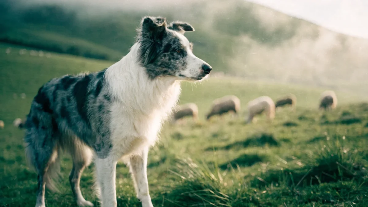
The border collie's gene pool tells a story of intense selective pressure aimed at a single outcome: the ability to herd sheep. For over a century, breeders chose mates based on working performance, not appearance. Color, coat type, eye shade — these were incidental. A dog that worked brilliantly was bred regardless of what it looked like. That approach created a breed with remarkable cognitive and physical capabilities, but it also carried forward genetic mutations with significant health implications.
Understanding border collie genetics matters most in three areas: the merle coat pattern and its risks, the inheritance of eye color, and the disease-causing mutations — particularly Collie Eye Anomaly — that DNA testing can identify and manage.
The Merle Pattern: Beautiful and Dangerous
Merle is the most visually striking coat pattern in border collies — a mottled patchwork of diluted and full-pigment areas that creates the blue merle (diluted black) and red merle (diluted chocolate) variations that many people find attractive. The pattern is caused by a SINE insertion in the PMEL17 gene (also called the SILV gene), which disrupts normal pigment distribution in individual hair follicles.
The merle gene follows an incomplete dominance pattern. A dog with one copy of the merle allele (Mm) displays the merle pattern with no associated health problems. The pattern is cosmetic — it affects appearance without compromising function.
The danger comes from double merle — a dog with two copies of the merle allele (MM). When two merle dogs are bred together, on average 25% of the puppies will be double merle. These dogs show excessive white coloring, often over 80% of the body and head, and carry a high risk of serious defects:
- Congenital deafness. Partial or complete hearing loss caused by degeneration of the cochlear structures. The risk correlates with the amount of white on the head, particularly around the ears.
- Ocular defects. Microphthalmia (abnormally small eyes), iris coloboma, and other structural eye abnormalities that range from cosmetic to functionally blinding.
- Combined sensory loss. Some double merle dogs are both deaf and blind, requiring specialized lifelong care.
Merle-to-merle breeding is avoidable with a single DNA test. Any breeder producing double merle puppies is either ignorant of the genetics or indifferent to the consequences. Neither is acceptable.
Cryptic Merle
A complicating factor in merle genetics is the existence of cryptic merle — dogs that carry the merle allele but display little or no visible merle patterning. A cryptic merle may appear to be a solid-colored dog and could be unknowingly bred to a merle partner, producing double merle offspring. This is why DNA testing for merle status is recommended before any breeding involving border collies, even when both parents appear solid-colored — a 2016 molecular survey of 500 Japanese border collies found the cryptic SINE-insertion allele at a frequency of 1.6%, low enough to hide easily in a pedigree until two carriers are paired.
Eye Color Genetics
Border collies display a wider range of eye colors than most breeds — from deep brown to amber, green, and blue. Heterochromia (two different-colored eyes) and sectoral heterochromia (two colors within a single iris) also occur, particularly in merle and piebald-patterned dogs.
Brown eyes are the most common and are produced by standard melanin deposition in the iris. Blue eyes in border collies are most frequently associated with the merle pattern — the same mechanism that dilutes coat pigment can dilute iris pigment, resulting in blue or partially blue eyes. However, blue eyes can also occur independently of merle through a separate, now formally identified mechanism: a duplication near the ALX4 gene on chromosome 18, first mapped in Siberian Huskies and since documented in border collies and several other breeds.
In non-merle border collies, blue eyes sometimes occur in dogs with significant white on the face, linked to the piebald (S locus) gene's effect on pigment distribution. Eye color in border collies has no effect on vision quality, regardless of shade — a blue-eyed border collie sees exactly as well as a brown-eyed one, assuming no other structural abnormality is present.
Collie Eye Anomaly (CEA)
CEA is the most important inherited eye condition in the breed. It is caused by a deletion in the NHEJ1 gene on chromosome 37 and follows autosomal recessive inheritance — a dog must carry two copies of the mutated gene (cea/cea) to be affected.
The condition affects the development of the choroid, a vascular layer at the back of the eye that supplies blood to the retina. The severity ranges enormously:
- Mild (Grade 1): Choroidal hypoplasia — a thinning of the choroid visible only on ophthalmoscopic examination. Dogs with mild CEA typically have normal functional vision throughout life.
- Moderate (Grade 2): Coloboma — a structural defect in the optic disc or surrounding tissue. May cause localized blind spots but generally does not lead to total vision loss.
- Severe (Grade 3-4): Retinal detachment or intraocular hemorrhage. Can cause significant vision loss or blindness. Relatively rare but devastating when it occurs.
A critical quirk of CEA diagnosis is the "go normal" phenomenon. Puppies with mild CEA examined before approximately 7-8 weeks of age show visible choroidal changes on ophthalmoscopy. After that age, the normal pigmentation of the retina may mask the lesion, making the dog appear ophthalmologically normal even though it carries and can transmit the condition. This is why DNA testing is more reliable than physical examination alone.
As explained in the breed's selective history, working selection prioritized herding ability rather than screening for eye conditions. CEA became widespread in the border collie population before DNA testing made carrier identification possible.
Managing CEA Through Breeding
With the DNA test, CEA is manageable: clear-to-clear pairings produce no affected offspring, clear-to-carrier pairings produce carriers but no affected dogs, and carrier-to-carrier pairings carry a 25% risk of affected puppies. The goal is never to produce affected puppies while gradually reducing carrier frequency — eliminating carriers too aggressively would narrow an already limited gene pool in some lines.
Other Testable Conditions
Beyond CEA and merle, border collies should be tested for Trapped Neutrophil Syndrome (TNS) — a fatal autosomal recessive immune disorder (VPS13B gene) where affected puppies rarely survive past 12 months — and MDR1 (ABCB1 gene), which causes dangerous drug sensitivity to ivermectin and other common medications. Neuronal Ceroid Lipofuscinosis (NCL), a rare but fatal neurodegenerative disease, also has a DNA test available. Details on all breed health conditions are covered in the border collie breed guide.
The border collie's gene pool is healthier than many purebred populations because working selection maintained a relatively broad base. However, popular sire concentration — a single outstanding trial dog siring hundreds of offspring — has created pockets of reduced diversity that breeders should monitor through genetic diversity testing.
Frequently Asked Questions
Is it safe to breed two merle border collies together?
No. Breeding merle to merle produces a 25% chance of double merle offspring, which carry serious risks of congenital deafness, vision defects, or both. This is entirely preventable by DNA testing merle status before breeding and never pairing two merle carriers. Any breeder producing double merle puppies is not following responsible breeding practices.
Can a border collie carry CEA without showing symptoms?
Yes. Border collies can be carriers (one copy of the mutation) with no clinical signs, or they can be mildly affected with changes detectable only by ophthalmoscopic exam — and after approximately 8 weeks of age, even mildly affected dogs may appear normal on eye exam due to the "go normal" phenomenon. DNA testing is the only reliable way to determine a dog's CEA status regardless of age.
Which genetic tests should border collie breeders run?
At minimum: CEA, TNS, MDR1, and merle status. Hip evaluation through a recognized screening program and annual eye examination by a veterinary ophthalmologist should supplement DNA testing. Additional tests for conditions like NCL, PRA, and degenerative myelopathy are recommended depending on breeding line history and regional prevalence.