

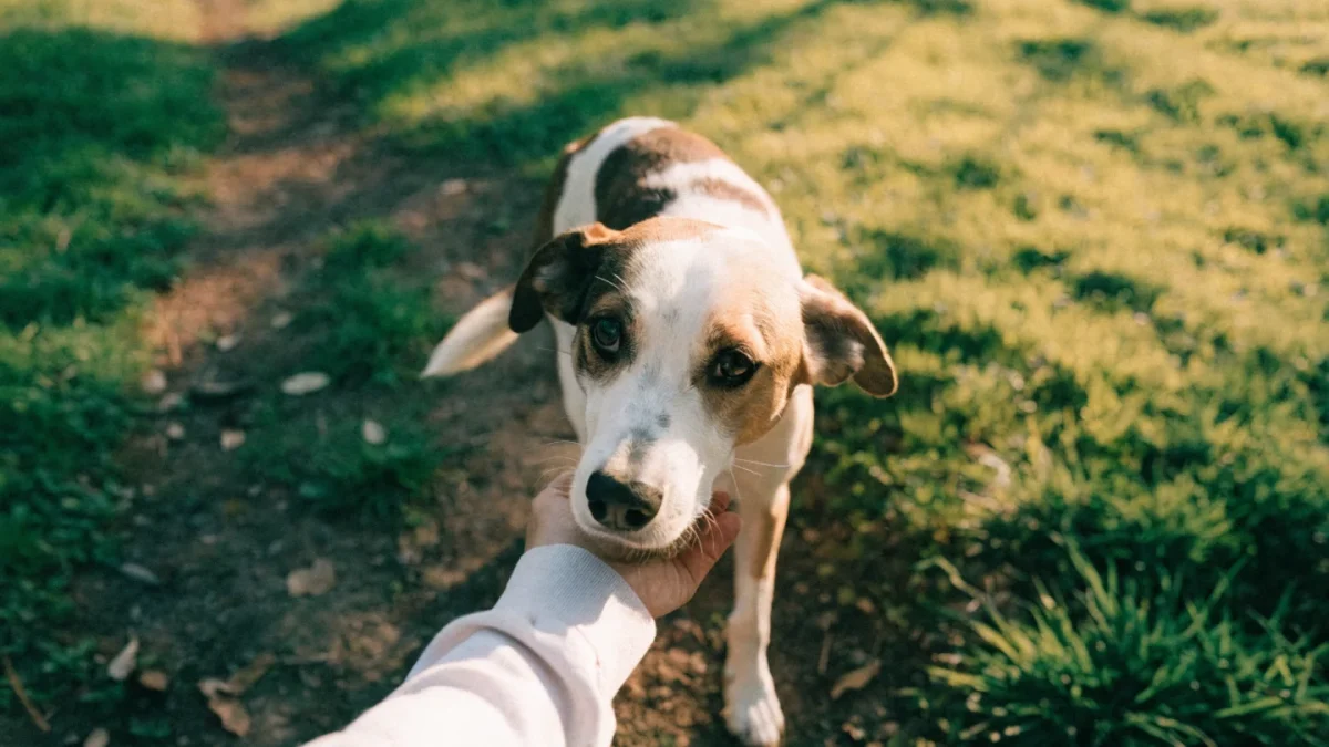
A terrier mix named Oscar came in one Saturday morning — the owner said he’d been running in the yard, yelped once, and was now carrying his back leg. Read our guide to joint and mobility health for detailed information. No obvious trauma, no fall, nothing he’d run into. He was holding the leg up completely without weight-bearing.
This presentation is one of the most common I see: a dog that was seemingly fine, then suddenly lame in a hind leg, usually after some degree of activity. In a dog like Oscar — seven years old, slightly overweight, not an athlete — the most likely diagnosis before I’d even palpated him was cruciate ligament disease.
The Landscape of Pet Joint Injuries
Joint injuries in pets fall broadly into several categories:
Ligament injuries: partial or complete tears of ligaments that stabilize joints. The cruciate ligaments (cranial and caudal, within the stifle/knee joint) are the most clinically important in dogs.
Sprains: overstretching or mild tearing of ligaments without complete rupture. Can occur in any joint, most commonly the carpus (wrist), tarsus (ankle), and stifle.
Joint luxations: displacement of one bone surface from another. Common in the patella (kneecap), hip, and elbow in dogs; in cats, hip and elbow luxations occur with trauma.
Meniscal injuries: damage to the cartilaginous menisci within the stifle joint, frequently occurring alongside CCL rupture.
Synovial injuries: inflammation and injury to the joint capsule from trauma or chronic instability.
Cranial Cruciate Ligament Disease: The Most Important
The cranial cruciate ligament (CCL) — anatomically equivalent to the anterior cruciate ligament (ACL) in humans — stabilizes the canine stifle (knee) by preventing excessive forward sliding of the tibia relative to the femur and internal rotation of the joint. When it fails, the stifle becomes unstable.
Why CCL Disease Is Mostly Degenerative, Not Traumatic
This is where the CCL differs fundamentally from the ACL. In people, ACL tears are overwhelmingly traumatic — sports injuries from sudden direction changes, contact, landing wrong. In dogs, most CCL ruptures are the end stage of a degenerative process that has been progressing for months or years.
The ligament becomes progressively weakened — fibers fray and fail — until a point where even normal activity causes complete rupture. The yelp Oscar’s owner heard wasn’t the ligament tearing from a trauma; it was the last fibers of an already compromised ligament finally giving way.
This has clinical implications: dogs with CCL disease often have changes in the other stifle too (because the same degenerative process is systemic), and they need management of the underlying condition, not just repair of the torn ligament.
What Makes Dogs Prone to CCL Disease
Several factors increase risk:
- Obesity: excess body weight strains all joint structures
- Breed: Labrador Retrievers, Rottweilers, Newfoundlands, Boxers, American Staffordshire Terriers, and Chesapeake Bay Retrievers have documented higher incidence; heritability of CCL rupture in Labrador Retrievers has been estimated at 0.62 — meaning approximately 62% of risk is genetic
- Tibial plateau angle (TPA): in dogs, the tibial plateau slopes posteriorly, creating a shear force that loads the CCL with every step. Dogs with steeper tibial plateaus experience greater CCL loading.
- Age: middle-aged to older dogs are at highest risk for degenerative CCL disease
- Neuter status: neutering is associated with increased CCL disease risk, particularly when performed before skeletal maturity. Multiple studies — including a 2024 JAVMA study and a large 4,000-dog survey of agility dogs — found that early gonadectomy significantly increases stifle injury risk, likely because sex hormones affect growth plate closure timing and ligament tissue maturation
Recognizing CCL Injury
Acute complete rupture (like Oscar):
- Sudden onset of hindlimb lameness, often non-weight-bearing or toe-touching only
- The dog typically holds the leg up
- May improve slightly over several days but won’t resolve without treatment
- Joint swelling develops
Partial tears and chronic disease:
- Intermittent hind limb lameness — may seem to improve, then worsen again
- Stiffness after rest
- Progressive muscle wasting over the affected limb
- Over time, joint thickening and arthritis development
The cranial drawer test — performed under sedation or anesthesia in most cases — assesses for abnormal forward movement of the tibia. A positive drawer sign confirms CCL rupture. The tibial compression test assesses for the same instability in a weight-bearing position.
Treatment Options
Conservative management (rest, anti-inflammatory medication, physiotherapy) is generally not considered adequate for dogs over 15–20 kg with complete CCL rupture. The joint instability is too significant, and OA progression is rapid without surgical stabilization. For small dogs under approximately 15 kg, conservative management has documented success rates of up to 85% in some series, and extra-capsular surgical outcomes are also excellent in that weight range; for cats, conservative management is often pursued with good results.
Surgical options for dogs:
Extracapsular repair (lateral suture/TightRope): places an artificial ligament outside the joint to provide stability while the body’s fibrous tissue reinforces. Technically simpler and lower cost than osteotomy procedures. Generally preferred for smaller dogs; less durable in larger, more active dogs.
Tibial Plateau Leveling Osteotomy (TPLO): cuts and rotates the tibial plateau to reduce the shear force that previously required the CCL for stabilization. After TPLO, the dog is stable without the ligament. Outcomes data is strong; the most commonly performed procedure for this condition in large dogs in many countries.
Tibial Tuberosity Advancement (TTA): advances the tibial tuberosity to change the patellar ligament angle, neutralizing the tibial shear force by a different mechanism than TPLO. Also widely used; comparative outcomes in a systematic review and meta-analysis (Frontiers in Veterinary Science, 2022) found both procedures had high long-term success rates, though TPLO and lateral suture had slightly superior subjective owner and veterinary assessments over TTA at 1+ years in some studies.
Recovery after CCL surgery is measured in months, not weeks. Controlled leash walking only for 6-8 weeks, progressive physiotherapy, then gradual return to normal activity. Full functional recovery takes 3-6 months.
Patellar Luxation
The patella (kneecap) sits in a groove — the trochlear groove — on the distal femur. When the groove is too shallow or the overall limb alignment is abnormal, the patella slips out of the groove. In small breeds, it typically luxates medially (inward); in large breeds, lateral luxation is more common.
Grades of patellar luxation:
- Grade I: patella can be luxated manually but returns spontaneously; rarely causes lameness
- Grade II: patella luxates spontaneously during activity; intermittent skipping or carrying of the leg
- Grade III: patella is most often out of the groove; persistent lameness
- Grade IV: permanent dislocation; inability to extend the stifle properly
Grades I and II in small breeds are often monitored conservatively — many affected dogs live comfortably with appropriate management. Grades III and IV, or any grade causing significant lameness, typically warrant surgical correction.
Hip Luxation
Hip luxation — where the femoral head pops out of the acetabulum — is usually traumatic (road traffic accidents, falls). Dogs present with sudden, severe non-weight-bearing lameness of a hindlimb with the leg in an abnormal position.
Reduction (putting the hip back in) can sometimes be performed under anesthesia as a closed procedure. If the hip re-luxates or the joint is too damaged for successful reduction, surgical options include open reduction and stabilization, or femoral head and neck ostectomy (FHO) as a salvage.
Sprains: The Underdiagnosed Injuries
Ligament sprains — stretch injuries without complete rupture — are common in active dogs. Carpus (wrist) and tarsus (ankle) sprains occur after jumps, falls, slipping, or rough play. Signs are:
- Lameness following activity, often improving with rest
- Swelling or heat around the joint
- Pain on direct palpation of the area
- Inconsistent — present after exertion, less apparent at rest
Diagnosis requires careful palpation and stress testing of ligamentous structures, sometimes X-rays to rule out fracture, and occasionally MRI for detailed soft tissue assessment.
Mild sprains respond to rest and controlled activity. Significant ligament injuries in weight-bearing joints — particularly the carpus — may need splinting or surgical stabilization.
Understanding arthritis that develops as a consequence of joint injury is important context: joint injuries that are inadequately treated inevitably accelerate OA development. Every ligament injury that leaves residual instability, every luxation with residual joint incongruity, and every injury with incomplete recovery becomes an accelerated source of cartilage damage. Treatment decisions affect the joint’s long-term health, not just the immediate problem.
Recovery and Rehabilitation Principles
Regardless of the specific injury, several principles guide recovery:
- Controlled rest in the early phase — not cage rest for months, but no uncontrolled activity
- Pain management — comfortable pets cooperate better with rehabilitation and are less likely to compensate in ways that create secondary injuries
- Gradual, progressive return to activity — typically weeks to months depending on severity and whether surgery was performed
- Physiotherapy and muscle building — essential for regaining stability; the muscles surrounding a joint are critical stabilizers
- Weight management — a heavier patient puts more force through recovering structures
- Swimming or underwater treadmill — allows exercise and muscle maintenance with minimal joint loading during recovery
For more on recognizing lameness and understanding why your dog is limping, including the range of causes beyond ligament injuries, that resource provides helpful context.
When to See Your Vet
Seek urgent assessment if:
- Your dog is non-weight-bearing or toe-touching on any limb following activity
- There’s obvious deformity, swelling, or crepitus (grinding) in a joint
- Sudden onset lameness that doesn’t resolve within 24 hours
Schedule an appointment if:
- Intermittent lameness that keeps recurring
- Reluctance to use a limb that comes and goes
- Muscle wasting in one limb compared to the other
Frequently Asked Questions
Is surgery always necessary for CCL rupture in dogs?
For dogs under roughly 15kg, conservative management with strict rest and physiotherapy has reasonable success rates. For larger dogs, conservative management alone rarely provides adequate long-term outcomes — the joint instability from CCL rupture in a heavier dog progresses to significant OA quickly without stabilization. An orthopedic evaluation with your vet or a specialist will guide the recommendation for your dog’s specific size, breed, and lifestyle.
How do I know if my dog has a sprain versus something more serious?
Both can cause lameness that may improve with rest. Features that suggest something more serious: non-weight-bearing rather than just limping, obvious joint swelling, pain that isn’t improving after 48-72 hours of rest, or recurring lameness in the same leg. Any lameness that doesn’t clearly improve with 1-2 days of rest deserves veterinary assessment.
Can cats tear their cruciate ligament?
Yes, though it’s far less common than in dogs, and when it does occur in cats, it’s more often traumatic (fall from height, road traffic accident) than degenerative. Recovery in cats can sometimes be achieved with conservative management, though surgical options exist for more severe cases.