

A Labrador Retriever named Margot came into my practice several years ago. Her owner had noticed she was "slowing down a bit" — taking longer to get up in the morning, reluctant to jump into the car. She was seven years old. When we evaluated her, she had bilateral hip osteoarthritis that was already quite advanced. With the right management, Margot lived comfortably for several more years — but those signs of slowing down had been present for at least eighteen months before anyone connected them to joint disease.
That gap between symptom onset and veterinary care is common, and it's one of the most significant factors in how well pets do with joint conditions. The earlier joint disease is identified and managed, the more options exist and the more of your pet's comfortable life is preserved.
Understanding How Joints Work
To understand joint disease, it helps to understand what a healthy joint looks like. In a normal synovial joint — the type that allows free movement at the hips, knees, elbows, and shoulders — several components work in concert:
Articular cartilage covers the ends of bones within the joint. It's smooth, low-friction, and has no nerve endings or blood supply of its own. This means cartilage can't hurt (it has no pain receptors), but it also can't heal itself effectively once damaged.
Synovial fluid lubricates the joint space, nourishes cartilage, and provides cushioning. In inflammatory joint disease, the composition of this fluid changes, contributing to pain and further joint damage.
The joint capsule surrounds and stabilizes the joint, containing the synovial fluid and providing structural support.
Periarticular structures — ligaments, tendons, and muscles — stabilize the joint and control its movement.
When any of these components are damaged — by developmental abnormalities, trauma, inflammatory disease, or simple wear over time — the joint becomes painful, less functional, and progressively worse if left unmanaged.
Types of Joint Disease in Pets
Osteoarthritis (Degenerative Joint Disease)
Osteoarthritis (OA) is by far the most common joint disease in both dogs and cats. It's a progressive condition characterized by the breakdown of articular cartilage, changes in the underlying bone, thickening and fibrosis of the joint capsule, and the development of osteophytes (bone spurs) at joint margins.
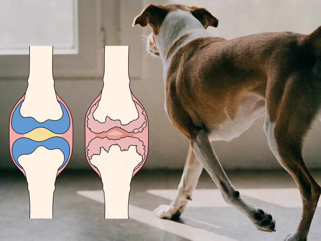
OA can be primary (occurring without an obvious preceding cause, more common in older animals) or secondary (developing as a consequence of another joint abnormality — like hip dysplasia, a previous ligament injury, or osteochondrosis). In practice, the distinction matters because addressing the underlying cause of secondary OA can slow its progression.
The condition is far more prevalent than most pet owners realize. Studies suggest that up to 80% of dogs over eight years old have radiographic evidence of OA in at least one joint, though not all are symptomatic — and a 2024 study specifically assessing four major joints in dogs over eight years found OA prevalence ranging from 36% to 57% depending on the joint, with the elbow most commonly affected. In cats, OA is similarly prevalent but dramatically underrecognized because cats are stoic and show pain in subtle ways that don't match dog-owner expectations.
Hip Dysplasia
Hip dysplasia, one of the most common developmental joint conditions in dogs, is a malformation of the hip joint where the ball (femoral head) doesn't fit properly in the socket (acetabulum). This loose, abnormal fit causes chronic inflammation, instability, and eventually secondary OA. Large and giant breeds are most affected, though it occurs across a wide spectrum of sizes.
Hip dysplasia has a strong genetic component, which is why breeding programs that screen and selectively breed against dysplastic conformation have reduced (though not eliminated) prevalence in some breeds.
Elbow Dysplasia
A group of developmental conditions affecting the elbow joint — including fragmented medial coronoid process, osteochondrosis of the humeral condyle, and ununited anconeal process. Like hip dysplasia, elbow dysplasia leads to progressive OA and is most common in large breeds. Diagnosis typically requires imaging (X-rays, CT scan) and sometimes arthroscopy.
Cruciate Ligament Disease
The cranial cruciate ligament (CCL) — the equivalent of the ACL in humans — is the most commonly injured ligament in dogs. Unlike the traumatic tears common in athletes, most canine CCL disease is a chronic, degenerative process where the ligament weakens progressively before eventually rupturing. The result is joint instability, pain, muscle wasting, and rapid development of OA.
CCL disease is described by the American College of Veterinary Surgeons as the most prevalent orthopedic problem in dogs, affecting an estimated 3–5% of dogs, and CCL repair is consistently the most common orthopedic surgery performed by veterinary surgeons in practice.
Inflammatory Arthritis
This category includes immune-mediated polyarthritis (where the immune system attacks joint tissue), infectious arthritis (from bacterial, fungal, or tick-borne infections), and erosive arthritis (where joint structures are progressively destroyed). Inflammatory arthritis is less common than OA but important to distinguish because treatment is fundamentally different — anti-inflammatory and immunosuppressive for immune-mediated forms, antimicrobial for infectious forms.
Patellar Luxation
Common in small breeds, patellar luxation occurs when the kneecap (patella) slides out of its normal groove. It can be congenital or acquired, and ranges from intermittent (the patella pops out occasionally and the dog shakes or carries the leg briefly) to severe (permanent dislocation with constant lameness). Many mild cases require no treatment; severe cases need surgical correction.
How Joint Disease Affects Daily Life
Joint pain profoundly affects quality of life in ways that go beyond visible limping. Understanding the subtle signs that your dog is experiencing joint pain is important because many dogs are remarkably stoic:
- Behavioral changes: grumpiness, snapping when touched in certain places, reluctance to interact
- Activity reduction: shorter walks, refusing to play, hanging back on terrain changes
- Stiffness: worse after rest, improves slightly with movement, worsens again after heavy activity (the classic OA pattern)
- Muscle wasting: visible loss of muscle mass over the hindquarters or shoulders as the animal offloads weight from painful limbs
- Altered posture: shifting weight, carrying a limb, a hunched or "tucked up" back
- Sleep disturbance: difficulty finding a comfortable position, changing positions frequently at night
Cats with joint disease show a particularly distinct pattern. You may not see limping at all. Instead, watch for:
- Reluctance or failure to jump to previously used heights
- Landing awkwardly or two-stepping down from heights
- Unkempt coat and reduced grooming, particularly over the back (painful to reach)
- Withdrawal from social interaction and favorite locations
- Changes in litter box use (difficulty climbing in or out)
- Increased aggression or irritability when handled in the hindquarters
Diagnosis: What Your Vet Is Looking For
Clinical Examination
A thorough orthopedic examination remains the foundation. Your vet will:
- Watch your pet move — gait analysis identifies which limbs are affected and how severely
- Palpate each joint — feeling for effusion (fluid), crepitus (grinding), pain response, and range of motion
- Assess muscle symmetry — muscle wasting over affected limbs is a reliable indicator of chronicity
- Perform specific orthopedic tests — such as the cranial drawer test for CCL rupture, or the Ortolani maneuver for hip laxity
Imaging
X-rays remain the first-line imaging tool for joint disease. They show:
- OA changes: joint space narrowing, osteophyte formation, subchondral bone changes
- Fractures and developmental abnormalities
- Hip dysplasia: shallow acetabula, abnormal femoral head shape
X-rays have limitations — they show bone changes but not soft tissue, and early cartilage damage isn't visible radiographically. CT scan provides superior bony detail, particularly for complex joints like the elbow. MRI is the gold standard for soft tissue assessment — cruciate ligament integrity, cartilage thickness, meniscal tears — but requires general anesthesia and is more resource-intensive.
Additional Diagnostics
Joint fluid analysis: aspiration and analysis of synovial fluid distinguishes OA (non-inflammatory, few cells) from inflammatory arthritis (high white cell count) from infection. Relatively straightforward, very informative.
Bloodwork and tick panels: important when systemic illness or tick-borne disease might be driving joint signs — polyarthritis from tick-borne infections can mimic primary OA.
Management: An Evidence-Based Approach
Joint disease in pets is managed, not cured. The goal is pain control, functional preservation, and slowing progression. A multimodal approach — using several strategies simultaneously — consistently outperforms single interventions, a position supported by the current consensus in veterinary orthopedic and pain management literature, which recognizes that the complex pathophysiology of OA cannot be adequately addressed by any single treatment.
Weight Management
This is consistently the single most impactful modifiable factor in joint disease. Every kilogram of excess body weight translates directly into additional force across load-bearing joints. Studies in dogs have demonstrated that weight loss alone significantly reduces lameness scores in obese dogs with OA — a peer-reviewed study published in Veterinary Research Communications (2010) found measurable improvement in lameness from as little as 6% body weight loss, with objective gait analysis improvement from 8.85% loss. Even in cats, reducing body condition to a lean score measurably improves mobility.
There's no medication, supplement, or surgery that compensates for an overweight body. Weight management should be the first conversation, not an afterthought.
Controlled Exercise
"Rest" is not typically the right recommendation for chronic joint disease. Controlled, low-impact, regular exercise maintains muscle mass (which supports and offloads joints), maintains joint fluid health, and prevents the stiffness and rapid deconditioning that comes with inactivity.
Appropriate exercise looks different across stages:
- Mild OA: regular leash walks, swimming or hydrotherapy if available, controlled play
- Moderate OA: shorter, more frequent sessions; avoid hard stops and starts, rough terrain; hydrotherapy is particularly valuable
- Severe OA or post-surgical: strict controlled movement per veterinary guidance; supervised physiotherapy
What's consistently inadvisable is high-impact, uncontrolled activity — long hikes followed by days of rest, vigorous fetch, stair-climbing in young large dogs at risk of developmental disease.
Pain Management
Effective pain control is both an ethical obligation and a practical necessity — pets in pain are inactive, which worsens muscle wasting and joint health.
NSAIDs (non-steroidal anti-inflammatory drugs) remain the first-line pharmaceutical intervention for OA pain in dogs. Newer formulations offer improved GI safety profiles. Regular monitoring (bloodwork every 6-12 months) is appropriate for long-term NSAID use to check liver and kidney function. NSAIDs in cats require extreme caution and veterinary supervision due to cats' unique metabolism.
Gabapentin and amantadine: used for chronic pain with a neuropathic component, often in combination with NSAIDs rather than instead of them.
Monoclonal antibodies targeting nerve growth factor (anti-NGF): bedinvetmab (dogs) and frunevetmab (cats) are licensed monthly injectable medications shown in randomized, placebo-controlled trials to provide effective OA pain relief and quality-of-life improvement. They offer a non-NSAID option for animals where NSAIDs are contraindicated or inadequate — and are described by some researchers as the most significant advance in veterinary OA pain management in 25 years.
Corticosteroids: used selectively, typically for inflammatory arthritis or as short-term bridges. Not recommended for long-term OA management due to effects on cartilage and metabolic health.
Physical Rehabilitation
Veterinary physical rehabilitation — physiotherapy for animals — includes therapeutic exercises, manual therapy, underwater treadmill hydrotherapy, therapeutic laser, and electrotherapy. Multiple published studies support its use: low-level laser therapy has demonstrated pain improvement and reduced analgesic requirements in arthritic dogs, and aquatic therapy (including underwater treadmill) improves joint range of motion and muscle mass in dogs with OA. Not universally available, but where accessible, it's genuinely valuable.
Joint Supplements
Glucosamine, chondroitin, and omega-3 fatty acids are commonly used supplements in pets with joint disease. The evidence is mixed for glucosamine and chondroitin, but marine-derived omega-3s (EPA and DHA) have the strongest evidence base: multiple randomized, controlled trials — including a double-blind multisite study in dogs — have shown significant improvements in lameness scores, weight-bearing, and reduced pain medication requirements compared to placebo. They're generally considered safe and worth discussing with your vet as part of a multimodal plan.
Environmental Modification
Simple home adjustments that reduce pain and improve daily quality of life:
- Orthopedic or memory foam beds — reduce pressure on sore joints
- Non-slip surfaces — rugs or mat runners prevent falls and the associated pain from compensatory muscle strain
- Ramps instead of stairs — for dogs who need to access furniture or vehicles
- Raised food and water bowls — reduce neck extension pain in dogs with cervical or shoulder OA
- Cat-specific: low-sided litter boxes, ramps to favorite resting spots, accessible food and water at multiple heights
Surgical Options
For certain conditions, surgery is the most effective management:
- Total hip replacement: highly effective for severe hip OA or end-stage hip dysplasia
- TPLO, TTA, or other CCL procedures: stabilize the stifle (knee) after CCL rupture
- Femoral head and neck ostectomy (FHO): a salvage procedure for severe hip disease when total replacement isn't possible
- Arthroscopy: minimally invasive joint surgery for specific indications
- Patellar groove replacement or realignment: for severe patellar luxation
Not every arthritic pet needs surgery, but for the right candidates with the right conditions, surgical intervention can be transformative.
The Difference Between Dogs and Cats
This deserves its own emphasis. Dogs limp. Cats don't — or rather, cats very rarely limp in a way owners recognize as lameness. A dog with a painful knee will likely show obvious changes in gait. A cat with bilateral hip OA may simply stop jumping to the windowsill, groom less thoroughly, and become slightly less interactive — changes that are easy to attribute to age or personality.
For detailed information on recognizing and managing joint disease specifically in these two species, see the dedicated guides on arthritis in dogs and cats — diagnosis, pain relief, and daily management.
Special Considerations for Young Pets
Developmental joint disease in young, growing animals is a separate consideration from age-related OA, though the two can coexist. Puppies of at-risk breeds benefit from:
- Appropriate nutrition: neither overfeeding nor underfeeding growing large-breed puppies; rapid growth exacerbates developmental joint disease. Excess calcium supplementation is particularly problematic — puppies cannot regulate intestinal calcium absorption the way adult dogs can, meaning excess dietary calcium is absorbed and associated with osteochondrosis, joint dysplasia, and other developmental orthopedic diseases. The recommended calcium range for large-breed puppies is 0.8–1.2% dry matter, with a calcium-to-phosphorus ratio between 1.1:1 and 1.4:1.
- Controlled exercise: avoid sustained, high-impact activity in large-breed puppies under 12-18 months; soft surfaces, leash walks, and limited stair use are appropriate
- Breeder screening: responsible breeders in high-risk breeds participate in joint health screening programs. Asking about parental hip and elbow scores is reasonable when acquiring a large-breed puppy.
Frequently Asked Questions
At what age should I start worrying about my dog's joints?
The answer depends on breed and size. Large and giant breeds are at higher risk for developmental joint disease that can manifest in the first 1-2 years of life, and for accelerated OA progression as early as 5-6 years. Small breeds often maintain good joint health longer. For any breed, baseline orthopedic evaluation is valuable in middle age (around 5-7 years), and sooner if there's any reason for concern.
Can cats really have arthritis?
Yes, and it's strikingly common. Studies using detailed radiographic evaluation of cats find OA in a very high proportion of cats over 10 years old. The misconception that cats don't get arthritis comes from the fact that they don't show it the way dogs do — no obvious limping, no vocalizing. The behavioral signs are subtler but real.
Is swimming good for dogs with arthritis?
For most dogs with OA, controlled hydrotherapy — either swimming or underwater treadmill — is excellent. The buoyancy of water dramatically reduces joint loading while allowing exercise that maintains muscle mass. It's particularly valuable after orthopedic surgery. Not every dog tolerates it, and it requires either an appropriate facility or safe water access, but for dogs that do well with it, hydrotherapy is one of the most beneficial interventions available.
Do joint supplements actually work?
The evidence for individual supplements is mixed. Omega-3 fatty acids (particularly EPA and DHA from marine sources) have the most consistent anti-inflammatory evidence and are reasonable to include. Glucosamine and chondroitin are widely used; some trials show benefit, some don't. The most honest answer is that they're unlikely to cause harm, may provide benefit for some animals, and should be considered part of a broader management plan rather than a standalone solution.
When does pain medication become necessary?
When a pet is showing behavioral or mobility changes consistent with pain — reluctance to move, reduced activity, stiffness, behavioral changes — pain management is appropriate. I prefer to manage pain proactively rather than waiting until a pet is visibly suffering. Untreated pain also drives muscle wasting, which worsens joint health in a feedback cycle. If you're unsure whether your pet is in pain, a veterinary assessment is the right first step.