

A 2012 study published in PLOS Genetics by Schoenebeck et al. first identified BMP3 as a gene strongly associated with skull shortening in small brachycephalic breeds including pugs. Then in 2017, a study published in Current Biology by Marchant et al. identified SMOC2 as a major additional contributor — a retrotransposon-mediated missplicing of SMOC2 that accounts for 36% of facial length variation in dogs tested, with pugs among the most extremely affected breeds. That finding gave a molecular name to something pug owners already knew: the flat face that defines the breed's appearance is not a single cosmetic feature. It's a skeletal reconfiguration with cascading effects on airways, eyes, dentition, and thermoregulation — all written in the DNA.
Understanding pug genetics brachycephaly PDE isn't just for breeders. It's essential knowledge for anyone living with or considering this breed, because the pug's genetic profile directly determines the health challenges its owner will navigate.
Brachycephaly: The Skull Gene
Brachycephaly in pugs results from a drastically shortened skull relative to width. The genetic architecture behind this involves multiple genes, but BMP3 (bone morphogenetic protein 3) carries the strongest effect. Variants in BMP3 reduce the length of the facial bones — the maxilla and nasal bones — without proportionally reducing the soft tissue structures that occupy the space.
This is the core problem. The soft palate, tongue, nasal turbinates, and other tissues that evolved to fit inside a normal-length muzzle are now compressed into a skull that's been shortened by centimeters. The soft palate elongates relative to the available space. The nares may be stenotic (abnormally narrow). The nasal turbinates can be distorted or overgrown, further obstructing airflow. The trachea may be hypoplastic (undersized for the body).
Collectively, these changes produce Brachycephalic Obstructive Airway Syndrome, which ranges from mild snoring during sleep to severe respiratory distress during minimal exertion. The degree of clinical expression depends on how many of these soft tissue changes are present and how severely each manifests — which is influenced by both genetics and individual development.
The pug breed guide discusses the practical health implications in detail. From a genetics perspective, the key point is that brachycephaly is polygenic and exists on a continuum. Pugs with slightly longer muzzles carry fewer or less extreme versions of the skull-shortening variants. This is why conformation choices in breeding programs directly affect respiratory outcomes in offspring.
Pug Dog Encephalitis (PDE)
PDE — formally known as necrotizing meningoencephalitis (NME) — is a breed-specific inflammatory brain disease that occurs almost exclusively in pugs. It causes progressive inflammation and necrosis (tissue death) in the brain, particularly in the cerebral hemispheres.
Symptoms typically appear between 9 months and 7 years of age and include seizures, circling, behavioral changes, blindness, and progressive neurological deterioration. PDE is ultimately fatal, with most affected dogs declining over weeks to months after symptom onset. There is no cure. Immunosuppressive therapy can slow progression in some cases but does not reverse the damage.
In 2011, researchers at the University of California, Davis — led by Dr. Niels Pedersen — published their identification of a strong genetic association between PDE susceptibility and the dog leukocyte antigen class II (DLA-II) region on chromosome 12 in the Journal of Veterinary Diagnostic Investigation. A commercial DNA test became available through the UC Davis Veterinary Genetics Laboratory that same year, priced at $55 per animal. The test identifies dogs as having a low, moderate, or high genetic risk for developing PDE based on their DLA-II haplotype.
The inheritance is complex — the genetic markers are associated with susceptibility rather than certainty. Not every high-risk dog develops PDE, and the environmental triggers that initiate the disease in genetically susceptible dogs are not yet fully understood. The autoimmune nature of the condition suggests that immune system dysregulation, possibly triggered by infection or other environmental factors, interacts with the genetic predisposition.
What the test does provide is risk stratification. Breeders can use DLA-II genotyping to avoid high-risk matings, and owners of high-risk dogs can work with their veterinarians to establish monitoring protocols. As the pug history article traces, the breed's narrow genetic base — a consequence of centuries of closed-population breeding — concentrated the susceptibility alleles that underlie PDE.
The Tail Curl: Hemivertebrae and Conformation
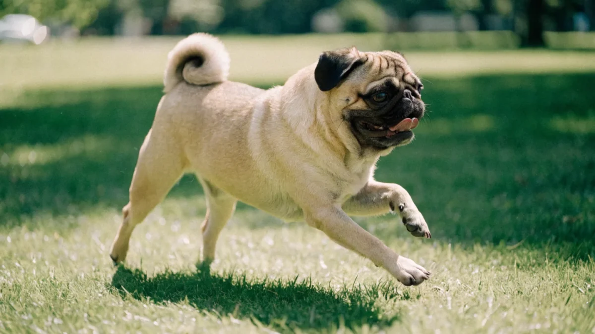
The pug's tightly curled tail — the double curl is considered ideal in many breed standards — is produced by hemivertebrae in the caudal (tail) vertebrae. Hemivertebrae are wedge-shaped vertebral bodies that cause the spine to curve rather than extend straight.
In the tail, hemivertebrae are cosmetically desirable and functionally inconsequential. The problem is that the same type of vertebral malformation can occur elsewhere in the spine — particularly in the thoracic region — where it causes spinal curvature (kyphosis or scoliosis), narrowing of the vertebral canal, and potential compression of the spinal cord.
Thoracic hemivertebrae are documented in pugs at significant rates — a 2017 study published in The Veterinary Journal found hemivertebrae in 17.6% of neurologically normal pugs, while a broader screening found that 96% of pugs presented with at least one congenital vertebral malformation of some type. Many affected dogs show no clinical signs, but severely affected individuals can develop progressive weakness, pain, and loss of coordination in the hind limbs. Radiographic screening can identify vertebral malformations before symptoms develop.
The genetic relationship between tail hemivertebrae and thoracic hemivertebrae is not fully resolved, but the association is strong enough that breeding for extreme tail curl may inadvertently select for increased spinal abnormalities elsewhere. This is a case where a conformation feature celebrated in the show ring carries a hidden cost to structural soundness. The Bulldog genetics article covering brachycephaly and health describes the same BMP3 and SMOC2 mechanisms at work in a closely related brachycephalic breed — the genetic architecture of flat-faced anatomy is remarkably consistent across breeds.
Coat Color Genetics
Pug coat color follows a relatively straightforward genetic pathway compared to some breeds, but it involves a few interactions worth understanding.
The two standard colors — fawn and black — are determined primarily by the A (agouti) and K (dominant black) loci. Black pugs carry at least one copy of the KB (dominant black) allele at the K locus, which overrides the agouti pattern and produces a uniformly black coat. Fawn pugs are ky/ky at the K locus, allowing the agouti locus to express, producing the characteristic fawn body color with a dark (melanistic) mask on the face.
The fawn shade ranges from light cream (sometimes called apricot) to a deeper reddish-gold. This variation is influenced by phaeomelanin intensity modifiers that are not yet fully mapped but clearly segregate within the breed. The dark mask and ear pigmentation are produced by the Em (melanistic mask) allele at the E (extension) locus — a dominant allele that concentrates eumelanin in the facial region.
Silver-fawn pugs — lighter, cooler-toned fawns — may carry dilution-related modifiers at the D locus, though the specific genetic mechanism producing the silver variant is not yet fully characterized, and some registries like the UK Kennel Club list silver as a distinct standard color while others do not. The practical significance is minimal for pet owners but matters for breeding programs tracking color outcomes.
Testing Priorities for Pug Owners
The pug's genetic profile creates a focused testing agenda:
- PDE risk assessment. The DLA-II test identifies susceptibility. High-risk dogs benefit from owner awareness and veterinary monitoring protocols.
- Spinal screening. Radiographic evaluation for hemivertebrae, particularly in breeding stock, identifies dogs with thoracic vertebral malformations that could produce clinical problems or be passed to offspring.
- Patellar evaluation. Patellar luxation has a genetic component in pugs and should be screened in breeding animals.
- Eye examination. The prominent pug eye is anatomically vulnerable to corneal ulceration and pigmentary keratitis. Ophthalmic evaluation establishes a baseline.
The pug's compressed genetic diversity — the product of thousands of years as a closed breed population — means that deleterious recessive alleles are more common than in breeds with wider gene pools. Testing isn't optional. It's how responsible ownership accounts for the genetic hand the breed has been dealt.