

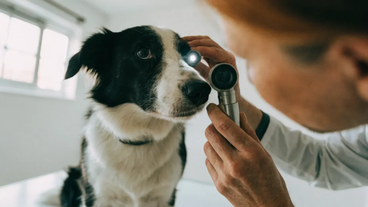
The eye is one of the more complex and delicate structures in the body — and for that reason, eye problems in pets can go from minor to serious faster than most other conditions. I've seen dogs lose vision from conditions that were eminently treatable, simply because the signs were not recognized or not taken seriously enough early on.
This guide covers the full range of common eye conditions in dogs and cats: what causes them, what they look like, and how they're managed.
Recognizing Eye Problems: General Signs
Before getting into specific conditions, it helps to know what to look for. Signs that something is wrong with a pet's eye include:
- Discharge: clear, watery, yellow, green, or brown
- Squinting or blepharospasm (holding the eye partially or fully closed)
- Redness in the visible white (sclera) or the conjunctiva
- Cloudiness in the cornea or within the pupil
- Visible swelling of the eyelids or third eyelid
- Asymmetry between the two eyes — size, appearance, pupil dilation
- Behavioral changes: bumping into things, reluctance to move in low light, rubbing the face
Any of these signs warrant a veterinary assessment. Eye conditions are among the presentations where I most strongly encourage owners not to wait.
Conjunctivitis
Conjunctivitis — inflammation of the conjunctiva — is the most common eye condition I see in both species.
In dogs, conjunctivitis is most often caused by bacterial infection, allergies, or as a secondary response to another eye problem (dry eye, entropion, corneal disease). Discharge is typically mucoid to mucopurulent (thick, yellow-green). Both eyes are frequently affected if the cause is systemic (allergies) but may be unilateral with localized infection or injury.
In cats, conjunctivitis has a different major cause: feline herpesvirus type 1 (FHV-1). This virus infects most cats at some point — often as kittens — and can remain latent, flaring during periods of stress or immunosuppression. Calicivirus and Chlamydophila felis are other causes — one study of cats with conjunctivitis found FHV-1 in about a third of cases versus roughly a fifth for Chlamydophila felis, making FHV-1 the more frequent single cause, though co-infections happen.
Treatment depends on the cause. Bacterial infections respond to topical antibiotic drops or ointment. Viral conjunctivitis in cats may be managed with antiviral agents. I used to recommend L-lysine supplementation routinely — it was standard advice for years — but I've stopped. Systematic reviews and current guidelines from the European Advisory Board on Cat Diseases found it doesn't meaningfully prevent or shorten flare-ups, so it's fallen out of favor in how I manage these cases now. Allergic conjunctivitis in dogs requires addressing the underlying allergy.
Corneal Ulcers
A corneal ulcer is a defect in the corneal epithelium — the surface layer of the eye. They range from superficial (affecting only the outer epithelium) to deep, stromal ulcers that threaten the structural integrity of the eye.
Causes include trauma (scratches from plants, cat claws, or self-trauma), foreign bodies, entropion (eyelids rolling inward), dry eye reducing corneal protection, and chemical irritation.
Signs are typically dramatic: intense squinting, excessive tearing, and obvious eye pain. The affected eye may appear cloudy. Pain is severe because the cornea is densely innervated.
Diagnosis is made by applying fluorescein dye — a green stain that binds to exposed corneal stroma but not to intact epithelium. Under a blue light, the ulcerated area stains bright green.
Treatment for superficial ulcers involves antibiotic eye drops to prevent secondary infection, pain management, and sometimes an Elizabethan collar to prevent self-trauma. Deep or non-healing ulcers require surgical intervention.
Brachycephalic breeds (Bulldogs, Pugs, Shih Tzus, Persians) are particularly vulnerable — their prominent eyes and reduced blink response make them prone to corneal exposure and ulceration. Any corneal ulcer in a brachycephalic dog is a more urgent situation.
Glaucoma
Glaucoma is elevated intraocular pressure (IOP) caused by impaired drainage of aqueous humor from the eye. The resulting pressure damages the optic nerve and retina, causing pain and, ultimately, blindness.
Primary glaucoma has a genetic basis in many breeds — Cocker Spaniels, Beagles, Basset Hounds, and others are predisposed, and researchers have even identified a susceptibility gene shared between some of these breeds and humans with glaucoma. Secondary glaucoma occurs as a consequence of other eye disease — uveitis (inflammation inside the eye), lens luxation, or advanced cataracts.
Acute glaucoma is an emergency. Signs include a suddenly painful, often hazy or cloudy-looking eye, redness, and behavior changes suggesting pain. The eye may appear enlarged. Vision loss can occur within hours.
Management includes medications to reduce IOP (both topical and systemic), and often surgery when medical management is insufficient. Once an eye has lost vision from glaucoma, the goal of treatment shifts to pain management and preventing the opposite eye from being affected.
For detailed information on glaucoma specifically, see the dedicated article in this cluster.
Cataracts
A cataract is an opacity of the lens — the transparent structure behind the pupil that focuses light onto the retina. It appears as a white or gray clouding visible within the pupil.
Causes include genetics (hereditary cataracts are common in many breeds including Golden Retrievers, Labrador Retrievers, and Boston Terriers), diabetes mellitus (diabetic cataracts in dogs develop rapidly and can cause complete blindness within days to weeks of diabetes onset — this is why newly diagnosed diabetic dogs need prompt ophthalmology assessment), advancing age, and less commonly inflammation or trauma.
Early cataracts may not significantly affect vision. As they progress, vision becomes impaired. Mature cataracts cause complete lens opacity.
Treatment is surgical — phacoemulsification (similar to human cataract surgery) uses ultrasound to break up and remove the lens, replacing it with an artificial implant. Outcomes are good in appropriate candidates. Not all dogs with cataracts are surgical candidates — age, systemic health, and retinal function all factor in.
Dry Eye (Keratoconjunctivitis Sicca — KCS)
KCS is a deficiency of the aqueous tear component, resulting in a dry, inadequately lubricated corneal surface. Without normal tear film, the cornea becomes irritated, thickened, and susceptible to secondary infections and ulceration.
Signs include a chronic mucoid discharge (often thick, green-gray, and sticky), a dull rather than bright corneal surface, squinting, and frequent eye infections. KCS is often confused with conjunctivitis because both produce discharge — the key difference is that KCS discharge is thick and the eye looks dry rather than watery.
Diagnosis is by the Schirmer Tear Test, a simple in-office test measuring tear production in mm/minute. Results below 15 mm/minute combined with clinical signs are considered suggestive of KCS; below 10 mm/minute with signs is generally enough to confirm the diagnosis, and below 5 mm/minute is considered diagnostic on its own.
Treatment is lifelong: cyclosporine or tacrolimus eye drops stimulate tear production in most dogs. These need to be applied one to two times daily indefinitely. Artificial tear lubricants supplement the medications. Most dogs with KCS are well managed on this protocol.
Cherry Eye
"Cherry eye" is the common term for prolapse of the third eyelid gland — the tear gland embedded in the third eyelid (nictitating membrane). When this gland loses its normal tissue attachment and prolapses, it appears as a round, red-pink mass in the inner corner of the eye, resembling a cherry.
It's most common in certain predisposed breeds — Bulldogs, Cocker Spaniels, Beagles, and Great Danes are frequently affected. It can occur in one or both eyes.
Treatment is surgical — the gland is repositioned and sutured back into place rather than removed (removing the gland permanently reduces tear production and increases the risk of KCS). Repositioning done by an experienced surgeon carries a success rate around 92%, and dogs who keep their gland carry a meaningfully lower lifetime dry eye risk than those who don't — though that risk is never zero.
Entropion and Ectropion
Entropion is inward rolling of the eyelid, causing the lashes and eyelid skin to rub against the corneal surface. This causes chronic irritation, corneal ulceration, and pain. It's common in breeds with loose facial skin — Chow Chows, Shar-Peis, Rottweilers, Mastiffs. Surgical correction is curative.
Ectropion is outward rolling of the lower eyelid, exposing the conjunctiva. It predisposes to conjunctivitis and foreign body accumulation. Common in Bloodhounds, Basset Hounds, and St. Bernards. Mild cases may need only supportive care; severe cases require surgery.
Progressive Retinal Atrophy (PRA) and SARDS
PRA is a group of inherited, degenerative retinal conditions causing progressive photoreceptor loss and eventual blindness. Early signs include night blindness and dilated pupils in low light. Most affected dogs progress to complete blindness. Genetic testing can identify carriers; no treatment exists.
Sudden Acquired Retinal Degeneration Syndrome (SARDS) causes rapid, permanent vision loss — often over days to weeks — in middle-aged dogs. The mechanism is not fully understood, despite decades of investigation — autoimmune and neuroendocrine theories are the leading hypotheses, and many affected dogs also show signs suggestive of Cushing's disease around the time vision is lost. Treatments aimed at those theories (high-dose steroids and other immune-modulating therapies) have been tried, but none has been shown in controlled studies to reverse or reliably prevent the vision loss.
Uveitis
Uveitis is inflammation of the uveal tract — the iris, ciliary body, and choroid. It presents as a red, painful eye with cloudiness (often a hazy or "steamy" appearance to the aqueous humor), altered pupil size, and sensitivity to light. It can be caused by infections, immune-mediated disease, trauma, lens disease, and cancer, and it predisposes to secondary glaucoma.
Uveitis requires prompt treatment with topical and often systemic anti-inflammatory medications to prevent secondary complications.
When to Act Immediately
Certain eye signs require same-day veterinary attention, not a "watch and wait" approach:
- Sudden cloudiness or haziness
- An eye appearing visibly larger than usual
- Any eye held tightly shut
- Visible injury to the eye surface
- A sudden change in apparent vision (pet is confused, bumping into things)
Many eye conditions deteriorate rapidly — just as ear problems in dogs and cats can escalate quickly if untreated. For most other health concerns, a 24-hour wait to see if things improve is reasonable — for eyes, it often isn't. For a complete overview of both eye and ear health in your pets, the pillar article on eye and ear health in dogs and cats covers prevention, inspection, and the broader context of these senses.
Frequently Asked Questions
Can a dog's eye infection clear up on its own?
Minor conjunctivitis from a transient irritant sometimes does. But most eye infections in pets don't resolve without appropriate treatment, and untreated infections can progress to deeper structures. If the eye looks red or there's discharge for more than 24 hours, a veterinary examination is appropriate rather than waiting.
Are eye conditions hereditary in dogs?
Many are. Cataracts, glaucoma, progressive retinal atrophy, entropion, and cherry eye all have strong breed predispositions with genetic components. If you have a breed known to be at risk for specific eye conditions, ask your vet about appropriate screening. Responsible breeders should test breeding stock for genetic eye conditions before breeding.
What does it mean if my cat's eye is squinting but not discharging?
Squinting without discharge is still painful — the cat is protecting a sensitive eye surface. Causes include corneal ulcers, uveitis, and early glaucoma, all of which need prompt assessment. For more on eye infections in cats specifically, the feline picture involves herpesvirus more prominently than the canine one.
How do I know if my pet is losing their vision?
Signs include: bumping into furniture or door frames, especially in low light; becoming hesitant in unfamiliar environments; startling more easily (can't see approach); looking "confused" in familiar spaces after layout changes; and in dogs with PRA, a characteristic shine to the eyes in darkness as the tapetum becomes more visible through deteriorating retinas. A simple "cotton ball test" — dropping a cotton ball near the pet and watching for visual tracking — can give a rough impression. Definitive assessment requires veterinary examination.